In recent years, machine learning has grown considerably and has proven to be useful in the medical image analysis field.
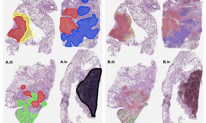
Recently, a team of research experts located at Dartmouth’s Norris Cotton Cancer Center leveraged machine learning abilities not only in overcoming the problem of grading tumor patterns but also the subtypes of lung adenocarcinoma, which is the most typical type of the primary cause of deaths resulting from cancer globally.
Today, lung adenocarcinoma requires a pathologist to carry out a visual examination of lobectomy slides in a bid to establish the tumor subtypes and patterns.
This classification plays a vital function in diagnosing and determining the best treatment for lung cancer, even though it is a subjective and difficult task.
SEE MORE: Artificial Intelligence in Medicine – Top Applications
The team, which was headed by Saeed Hassanpour, used the latest machine learning advances to build a deep neural network in classifying the various forms of lung adenocarcinoma, particularly on histopathology slides and discovered that the model was able to perform on the same level with three pathologists.
“Our study demonstrates that machine learning can achieve high performance on a challenging image classification task and has the potential to be an asset to lung cancer management,” says Hassanpour.
“Clinical implementation of our system would be able to assist pathologists for accurate classification of lung cancer subtypes, which is critical for prognosis and treatment.”
The team’s findings, “Pathologist-level classification of histologic patterns on resected lung adenocarcinoma slides with deep neural networks” are featured in Scientific Reports.
SEE MORE: Top 10 Ways Artificial Intelligence is Impacting Healthcare
Upon realizing that the technique could be applied to other numerous histopathology image analysis efforts, Saeed Hassanpour and his team availed their code publicly in an effort to promote new collaborations and research in the particular domain.
Apart from testing the performance of the deep learning model in a clinical environment to authenticate its potential to boost the classification of lung cancer, the team intends to apply the approach to other demanding histopathology image analysis activities in colorectal, breast, and esophageal cancer.
“If validated through clinical trials, our neural network model can potentially be implemented in clinical practice to assist pathologists,” says Hassanpour.
“Our machine learning method is also fast and can process a slide in less than one minute, so it could help triage patients before examination by physicians and potentially greatly assist pathologists in the visual examination of slides.”
Lung adenocarcinoma entails a type of non-small cell lung cancer that typically develops in tiny airways like bronchioles, and is commonly situated along the lungs’ external edges.
The condition cannot be eradicated entirely through surgery.
Like other lung cancer stages, the treatment of this condition relies on the overall health of the patient.
Adenocarcinoma begins in the cells located in the glands and can be diagnosed using different tests to establish whether it has spread to other body parts.
Some of the tests and steps utilized in diagnosing this subtype of lung cancer include biopsies, laboratory tests, and imaging tests.
Nevertheless, these techniques are applied to a person based on his or her location of the nodule(s), symptoms, medical condition and history, and other test results.
Source: MedicalExpress