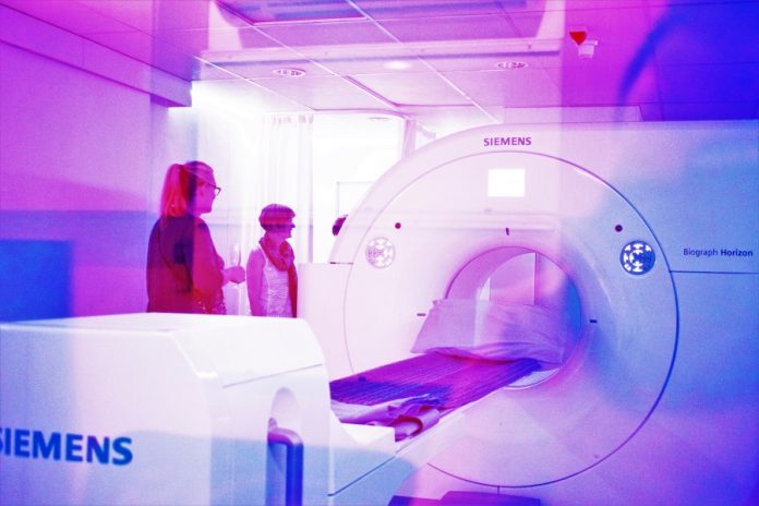
It’s good news all around for potential brain haemorrhage patients, thanks to a new artificial intelligence (AI) software solution that’s been developed by ParallelDots Inc. The application is called RADnet and the way in which it works is by detecting brain haemorrhages from CT scans.
Rather than look to replace the role of doctors, RADnet has been developed to act as alongside them as a second opinion. In doing this, the company is confident diagnosis times can be reduced, and survival rates can increase. “It’s important for the radiologist to know about a critical case as soon as it comes,” says co-founder of ParallelDots, Angam Parashar.
“It allows them to save time and provide a faster diagnosis to the patient. AI can help radiologists in making accidental findings in medical images.” The usual methods for detecting hemorrhages are often very time consuming and need a radiologist to be present throughout the whole process. Using the new AI software, that would all change.
The software was developed using deep neural networks to obtain and analyze anonymous medical data. Medical professionals then tidied up the data in preparation for the machine learning part of the process. A convolutional neural network called DenseNet was then used to model each CT scan in 2D. While that network focused on the haemorrhage regions, a bidirectional long short-term memory (LSTM) network was used to model the CT scan in 3D.
READ MORE – Top 10 Ways Artificial Intelligence is Impacting Healthcare
READ MORE – Artificial Intelligence in Medicine – Top 10 Applications
In comparing RADnet’s performance to that of three human radiologists, it scored higher in recall than two of them and the same as the third; it had higher F1 scores than two of the radiologists and it had the same accuracy level as one. “One big challenge that we see is around the performance of the AI system,” says Parashar. “We are dealing with critical cases here.
The AI technology needs to be really accurate to be meaningful to a radiologist. Additionally, it is important to consider other clinical parameters when developing the AI system. Combining all the information with medical imaging data is another challenge that needs to be addressed.”
So far the AI model has only been trained on 205 different CT scans, so is still quite limited in what it knows. Even so, the accuracy in which it performed against human radiologists provides hope that someday soon it will be a system that’s much more widely developed and able to be used in hospitals around the world.
Source Dotmed
